Lower Leg Bone Diagram Labeled - 16 best Bones in the Leg images on Pinterest | Human body ... : Below given knee diagram will help you to understand.. Below given knee diagram will help you to understand. Almost every movement in the body is the outcome of muscle contraction. Foot and ankle diagram anatomy. Label number 1 in the diagram indicates which part of the bone. System diagram labeled 209 human muscular system diagram labeled.
The tibia, or shin bone, spans the lower leg, articulating proximally with the femur and patella at the knee joint, and distally with the tarsal bones, to form the ankle joint. 3d rendering illustration of skeleton bone anatomy. Below given knee diagram will help you to understand. Lower leg muscle diagram blank sketch coloring page. Download a free preview or high quality adobe illustrator ai, eps, pdf and high resolution jpeg versions.
Bones of the Lower Limb | Anatomy and Physiology from s3-us-west-2.amazonaws.com When you stand or walk, all the weight of your upper body rests on them. System diagram labeled 209 human muscular system diagram labeled. Below given knee diagram will help you to understand. For more detail of the human bone structure, please visit: Below given knee diagram will. File human arm bones diagram svg labeled skeletal system diagram human leg bone structure They support the legs to bear the body weight and also help any disorder or defect in the knee bone can restrict the activities of the leg which can directly affect our locomotion. Learning to communicate nursing care:
Case history drug therapy laboratory diagnostics check your progress:
The tibia, or shin bone, spans the lower leg, articulating proximally with the femur and patella at the knee joint, and distally with the tarsal bones, to form the ankle joint. Leg muscle anatomical structure, labeled front, side and back view diagrams. Descriptionhuman leg bones labeled ru.png. Anterior view with primary bones names. They connect the lower leg to the rest of the body and gives stability, flexibility and strength. Your leg bones are the longest and strongest bones in your body. Unit 3 part 1 x section bone. File human arm bones diagram svg labeled skeletal system diagram human leg bone structure The foot bones shown in this diagram are the talus, navicular, cuneiform, cuboid, metatarsals and calcaneus. Vector illustration with human skeleton scheme isolated on a white background. The two bones beneath your knee that make up your shin are your tibia and fibula. Lower leg muscle diagram blank sketch coloring page. Label number 1 in the diagram indicates which part of the bone.
Vector illustration with human skeleton scheme isolated on a white background. The covering of a bone. System diagram labeled 209 human muscular system diagram labeled. Descriptionhuman leg bones labeled ru.png. Learning to communicate nursing care:
leg muscles labeled | A&P.2.Skin.Bone.Muscle | Pinterest ... from s-media-cache-ak0.pinimg.com Vector illustration with human skeleton scheme isolated on a white background. Blood and bones kids drawings. The foot bones shown in this diagram are the talus, navicular, cuneiform, cuboid, metatarsals and calcaneus. Master leg and knee anatomy using our topic page. Unit 3 part 1 x section bone. It widens and forms two condyles. Learn vocabulary, terms and more with flashcards, games and other study tools. Proximally, there are five key features of the tibia:
Foot and ankle diagram anatomy.
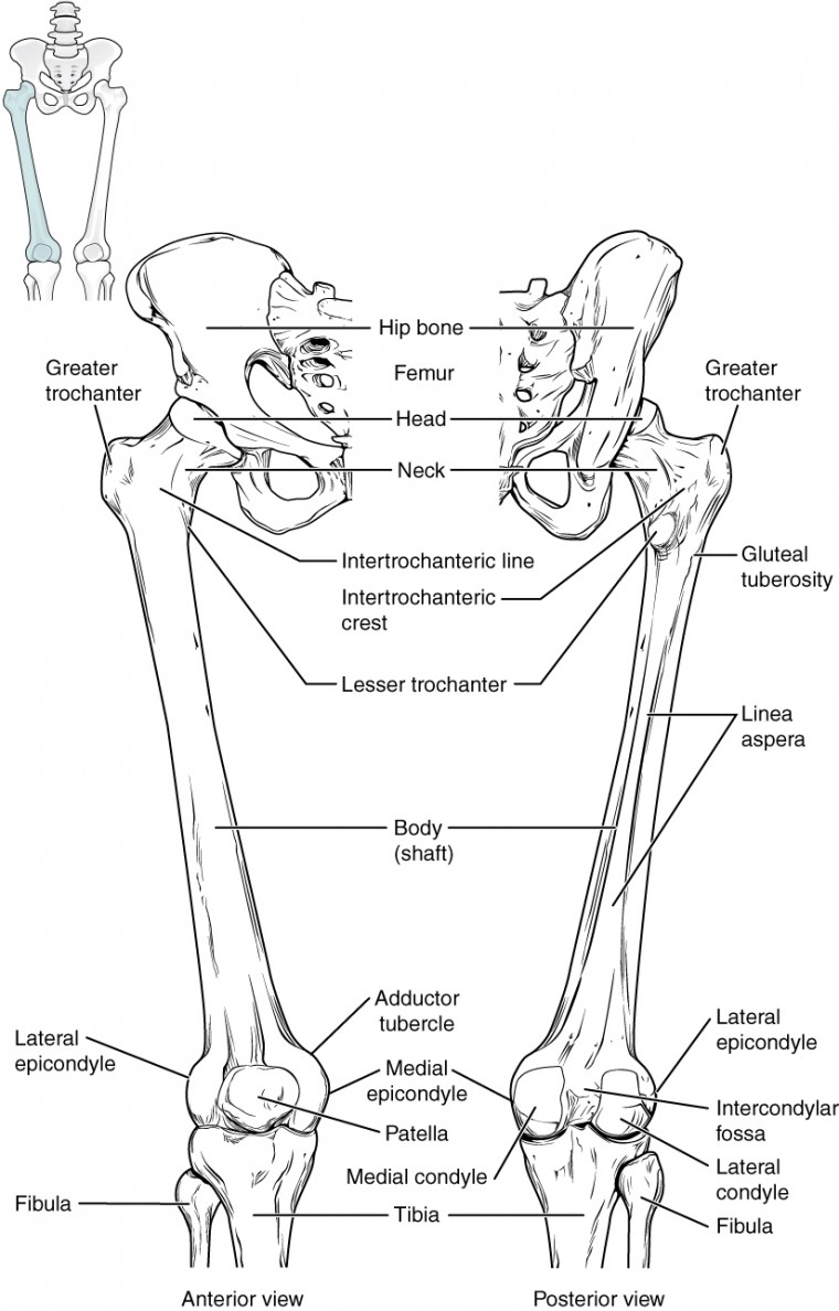
Download a free preview or high quality adobe illustrator ai, eps, pdf and high resolution jpeg versions. Lower bones limbs limb leg diagram muscle foot template anatomy blank human skeleton coloring sketch function th. Learning to communicate nursing care: The bones of the leg are the femur, tibia, fibula and patella. Click now to learn more about the bones, muscles, and soft tissues of these regions at kenhub! Long bones are longer than they are wide and are the major bones of the limbs. Proximally, there are five key features of the tibia: File human arm bones diagram svg labeled skeletal system diagram human leg bone structure Foot and ankle diagram anatomy. Descriptionhuman leg bones labeled ru.png. Vector illustration with human skeleton scheme isolated on a white background. The legs of the stick figure point to rounded projections located at the distal end of the femur known. Below given knee diagram will help you to understand.
The lower leg is comprised of two bones, the tibia and the smaller fibula. Leg muscle anatomical structure, labeled front, side and back view diagrams. Your leg bones are the longest and strongest bones in your body. Label number 1 in the diagram indicates which part of the bone. Learn vocabulary, terms and more with flashcards, games and other study tools.
Epicondyle of the knee from www.anatomynote.com Master leg and knee anatomy using our topic page. Your upper and lower leg are connected by a hinge joint. Leg muscle anatomical structure, labeled front, side and back view diagrams. The knee joint is the largest joint in the body and is primarily a hinge joint, although some sliding and rotation occur. For more detail of the human bone structure, please visit: Related posts of bone anatomy lower leg. 16 best bones in the leg images on pinterest. Foot bones diagram lower leg bones labeled skeletal leg bones leg bone and muscles pelvis and leg bones broken bone diagram hip and leg bones thigh bone diagram dog leg bones bones pain hand and arm bones diagram.
Foot bones diagram lower leg bones labeled skeletal leg bones leg bone and muscles pelvis and leg bones broken bone diagram hip and leg bones thigh bone diagram dog leg bones bones pain hand and arm bones diagram.
Case history drug therapy laboratory diagnostics check your progress: Blood and bones kids drawings. Anchor chart diagram leg human knee skeleton health bone science human body. Leg bones diagram unlabeled : Bones of the human lower limb. File human arm bones diagram svg labeled skeletal system diagram human leg bone structure The foot bones shown in this diagram are the talus, navicular, cuneiform, cuboid, metatarsals and calcaneus. Below given knee diagram will help you to understand. Femur bone anatomy made easy using a labeled diagram of the main parts of the thigh bone along with includes a blank diagram at the end to label on your own and quiz yourself! Long bones are longer than they are wide and are the major bones of the limbs. Muscles, connected to bones or internal organs and blood vessels, are in charge for movement. Related posts of bone anatomy lower leg. For more detail of the human bone structure, please visit:
Related posts of bone anatomy lower leg leg bone diagram. Related posts of bone anatomy lower leg.


0 Komentar